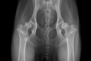
 Canine hip dysplasia (CHD) is arguably the most common orthopaedic disease seen in dogs. It is a degenerative condition where one or both of a dog’s hip joints do not develop normally. Affected dogs have an abnormally shaped “ball and socket” hip joint such that that “ball” of the hip does not sit tightly inside the “socket” part. In addition, the ligaments and soft tissue structures that hold the joint tightly together are abnormally loose. This looseness or laxity allows for excessive movement within the joint, resulting in the hip joint actually dislocating as the dog walks. This abnormal development and laxity in the joint results in excessive wear on the joint cartilage eventually leading to the development of painful arthritis.
Hip dysplasia is passed from parents to offspring through the genes. It is known to be most common in pure-bred large breed dogs such as German shepherds, golden retrievers, Labrador retrievers and Rottweilers. However, it can also occur any breed of dog (including cross-breeds) and of any size. It is also seen in some breeds of cats such as the Maine coon.
What is PennHip?
In recent years, a new diagnostic test to detect the likelihood of a dog developing hip dysplasia has been developed called PennHip. PennHip is a novel method to assess, measure and interpret the degree of laxity or looseness in a dog’s hips. Research has shown that the looser a dog’s hips are, the greater the chances it has of developing CHD later in life.
The PennHip assessment process
In order for PennHip to be of use it consists of three components:
Firstly, three radiographs of the dog’s hips are taken. The dog must be anaesthetised or heavily sedated as most dogs will not lie in the correct position or tolerate the manipulation that must be performed to obtain the images. Three different angles of the x-ray are taken to get a thorough understanding of the dog’s hip condition, including the presence of any arthritis that may have already developed.
Secondly, the radiographs are analysed by vets. Only vets who have completed specific training and certification are able to conduct the analysis. At Karingal Veterinary Hospital we have several veterinarians that are PennHip trained and certified.
 Thirdly, the x-rays are digitally submitted to California, USA and compared to a comprehensive database consisting of tens of thousands of results from dogs all around the world. A mathematical equation and analysis software is applied to the x-ray images to determine a Distraction Index (DI) Score. This score is a value between 0 and 1 and gives an indication of how loose the hips are. The closer the score to 0 the tighter the hips are and the less likely that the dog will go on and develop hip dysplasia. The higher the DI score the looser the hips. As an example, a DI of 0.5 means that 50% of the ball part of the hip joint exits the socket when the distraction view is taken. A DI of 0.75 means that the ball part comes out 75% of the way and it is 25% more loose than the dog scoring 0.5. As such it is much more likely to develop hip dysplasia.
Each dog submitted for PennHip receives a report, normally within two or three days. This report shows the DI score for each hip and any remarks from the radiologist who examined the x-ray images. The report also shows gives a Laxity Profile Ranking. This sections shows what the average DI score is for dogs of the same breed and gives an indication of where the individual dog lies with respect to all other dogs of the same breed. A sample PennHip report can be viewed here.
Why Do PennHip?
There are two very important reasons to perform PennHip radiographs on a dog. These are:
For much more information regarding PennHip please visit the Antech Imaging Services PennHip website here.